iMolview is an app for the iPhone/iPad and Android that lets you browse protein, DNA, and drug molecules in 3D. The app has a direct link to the Protein Data Bank (PDB) and DrugBank
and has a fast and easy to use interface. Touching the molecules via the screen allows you to interact immediately with the 3D structures in a unique way. You can zoom in and out, rotate, spin, pan, and clip
the 3D molecules with your finger tips in ways that are impossible using a traditional mouse and desktop computer.
Download
iMolview can be downloaded directly onto your iPhone or iPad by visiting the Apple App
Store. or onto your Android device Google Play Store.
iMolView allows you to search for drugs such as 'ibuprofen' or 'gefitinib' in DrugBank, or proteins like 'insulin' or 'thyroid receptor' in the
PDB and immediately explore their structure and annotation. This handy app allows you to quickly and easily view and interact with 3D molecules anywhere and at any time whether you are at the bench, at a conference, or just having coffee with colleagues!
The key features are:
Easy to use touch interface.
Save structure and slides (molecular document).
A direct link to the Protein Data Bank (PDB) and DrugBank.
Structure files (pdb/icb) sync with iTunes.
Supports retina display
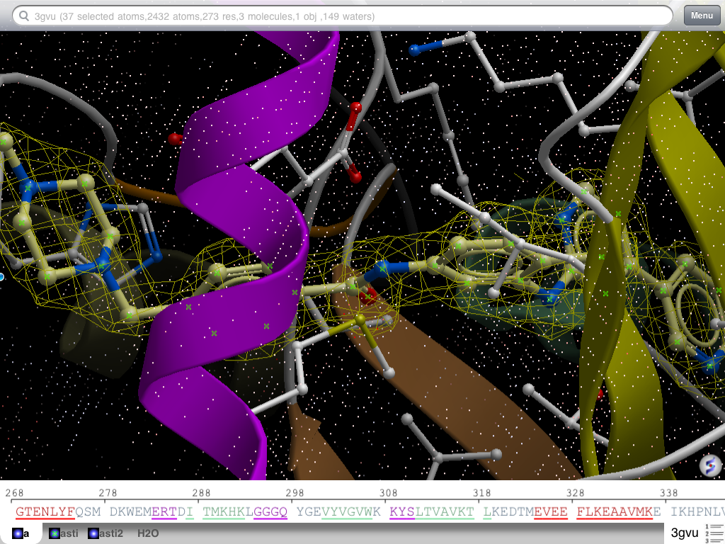
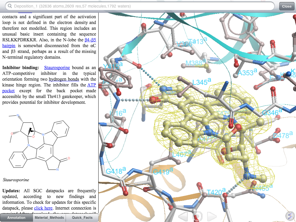
Display and contour electron density maps
Superimpose multiple objects
Side by side stereo
Angle and distance measurement
Each molecular view can be customized with a rich set of molecular representations including: wires, balls-and-sticks, space filling, ribbon diagrams, and molecular surfaces.
Zoom in and out, rotate, spin, pan, and clip the 3D molecule.
Search and download structures from NCBI website.
Read in any PDB file from any URL.
A wide selection of coloring schemes is available.
Color background, color molecule by atom type, chain, N- to C- terminal, and secondary structure.
Select residues, atoms, or chains and color or change their representations individually.
Select residues in sequence and get the corresponding selection in 3D.
Select the whole chain by holding the corresponding tab.
Display fog effect.
Set 'inertia' to the maximum and let your molecule spin in 3D indefinitely.
Display residue and site labels for the whole object or selection.
Direct link to PubMed, Uniprot, and RCSB.
Read in your own MolSoft ICM *.icb files.
Release Notes
Note: iMolview is under active development and is frequently updated with new features and bug fixes.
Version 1.7
Added 'Tools/Load Electron Density Map' to download crystallographic data for the currtently loaded object
Toggle map display in 'Misc' menu tools
Version 1.6
Added interface to Dropbox (Documents/Dropbox)
Added molecular skin/surface representation
Added "Tools/Filter Selection" to adjust the current selection by various parameters
"Color Selection" can color individual representations.
ICM scripts embedded with the ICB file are available in the menu.
Added full screen mode.
Added interface to the ICM command language (Command tab in the search bar)
Tools menu (sbs stereo, measurements) appears for small molecules
2D labels are displayed properly
Version 1.5
Major:
retina (high res) display support for the new iPad
multiple object support + superimpose
Others:
assign secondary structure
images in html pane
side-by-side stereo
angle and distance measurement
email screen shot
Version 1.4
added auto 'rocking&rotation mode'.
custom URL scheme imolview:// is extended to allow open document on the particular slide (from the webpager, keynote or other apps)
Added the ability to save molecular document (structure + slides).
Added the ability to save view and representation (slides).
Fixed memory leak in xstick display
Fixed crash in 3D labels
Version 1.3
support for horizontal screen orientation
support for custom URL scheme (imolview://)
search results are now scrollable and load more hits
VGA output on iPad and iPhone4 (3D view is mirrored, 'laser pointer' appears on touch and hold)
improved selection in sequence view (residue range is shown). Fixed bug with selecting single residue.
added atom labels display
added centering without zoom (via center button in menu)
preferences are now persistent even if the app is closed and opened again
added preference for hydrogen display (none/polar/all)
added default url preference for loading molecules off a web page
Version 1.2
issue with older iPhone/iPod devices FIXED
structure files (pdb/icb) sync via iTunes
download history
easy toggle of individual molecule display via sequence view tabs
faster refresh in CPK representation
display tools popup bar on selection
improved handling of very large structures
neighbors in other chains selection, such as atoms in the vicinity of selected ligand
external links menu: Uniprot and RCSB entries in addition to PubMed
atom type coloring for hetero-atoms only - allows combining chain-specific colors and atom type color coding
3D residue picking is now tracked in sequence view
easy clear display button (X)
bug/crashes fixes
iMolview Help
How to Display PDB Files
Here are some ways to read in PDB files into iMolview:
1. Enter PDB Code in the Search Panel
The standard way to read in PDB files is to simply type in the PDB code in the search panel at the top of the screen.
2. From any Website
If your PDB file is saved on a web server (at any URL) you can download it by going to the Menu button and selecting Load File(icb/pdb) From The Web.
In the View or Save 3D structure panel select 'PDB' instead of Cn34D and click on view structure. You'll be prompted to open it in iMolview.
Click OK.
Load your PDB/ICB Files via iTunes
Connect your iPhone or iPad to iTunes.
Drop your PDB or ICB file into File Sharing Area in iTunes (see image below).
Sync your iPad/iPhone with iTunes
Disconnect device and start iMolview. Go to Menu/Documents and your uploaded files will be listed there.
Click to enlarge image.
Navigation in the 3D View of the Molecule
Rotate
Touch with one finger and drag it in the appropriate direction. You can also let molecule spin the by flicking the screen quickly.
Adjust 'Inertia' parameter in the 'Settings' section to let the molecule spin longer or indefinitely
Zoom in/out
Touch the screen with two fingers and move them apart/together.
Z-Rotate
Touch the screen with two fingers and twist in the appropriate direction.
Pan
Touch the screen with two fingers and move them in any direction.
Clipping
Touch and move one finger on the right margin. Touching in the top/bottom half initiates movement of the back/from clipping plane. Two finger touch on the right margin initiates slab movement (back and front plane simultaneously).
Select
Touch and hold one finger at an atom/residue until floating label balloon appears. Tap on the balloon to select.
Cancel selection
Double tap anywhere in the 3D view.
Entire molecules/chains can also be selected by touching and holding on the bottom tab bar.
Sequence segments selections are done by touching/holding on the sequence view. Once selection is initiated,
beginning/end flags appear and can be dragged to extend or shrink selected sequence segment.
Selections are visually represented in 3D view as green crosses floating on selected atoms.
While the selection is active, tapping on the 'target' icon in the bottom of the view centers on selected molecules/residues/atoms.
Also, display representations and coloring are applied to the selection.
Saving Slides and Documents
Slide is a combination of a view point and graphics representation. In iMolview you can create multiple slides and access them later
after the document was saved.
To create a slide:
Setup a view and representations (color, label, etc.)
Go to Menu/Slides and choose "Save Current View"
Give a name and press 'OK'. It should appear in the list
Repeat these steps to create more slides
To modify or delete an existing slide hold your finger over the appropriate item. The possible actions are:
Rename: Rename the slide
Overwrite : overwrite the slide with the current view
Delete : delete the slide
To switch between slides you may use Menu/Slides or arrows on the sides of the center button.
After all slides are done you need to save your molecular document. Goto Menu/Documents/Save Current Document and give it a name.
The document will be saved into History folder and can be loaded later. This file can also be copied to your PC,MAC or Linux and
opened with free ICM-Browser, or you can embed this document as a live 3D object into
web page using ActiveICM technology.
Displaying Electron Density
Tools/Load Electron Density Map
Loads electron density map from Uppsala EDS server (http://eds.mbc.uu.se/eds/). Once map is loaded you can toggle its display using 'Misc' panel in the main menu. In addition map can be contoured around selected atoms:
Select part of the molecule
Click on the contour icon in the 'Misc' panel on the pain menu.
Contouring level can be adjust using slider in the 'Tools' menu.
New icons in 'Misc' panel:
Map view toggle: [Optional] toggles display of the electron density map (if loaded). (See Tools sections to see how to load a map)
Map Contour toggle: [Optional] Contour density map around selected atoms. Contouring level can be adjusted in the Tools menu.
Using custom URL scheme imolview://
Custom URL scheme can be used to launch iMolview from different applications (Safari, Keynote and others).
It has the following format:
imolview://[][?PARAM=VALUE&PARAM=VALUE]
PATH_TO_THE_FILE can be either relative local path to the Documents folder. (History/3gvu.icb) or absolute URL (http://side.com/folder/file.icb)
Supported file types are:
ICB: Molsoft molecular document format. May contains multiple molecular objects, meshes slides and others.
PDB: Protein Data Bank file.
MOL/MOL2: MDL or Tripos small molecule file.
PARAM can be one the following:
BGCOLOR: redefine the background color. The VALUE should be a color name.
PDBCODE: loads PDB structure with the code from VALUE. If you have this field PATH_TO_THE_FILE should be omitted.
SLIDE: sets the slide. The VALUE should be either slide name or number
RUN: runs a list of semicolon (or new line) separated lits of ICM script commands.
Examples:
# opens the document and sets the default view.
imolview://History/3gvu.icb
# opens slide 1
imolview://History/3gvu.icb?SLIDE=1
# opens slide 'Slide2'
imolview://History/3gvu.icb?SLIDE=Slide2
# opens default view for PDB '1xbb' sets background color to white
imolview://?PDBCODE=1xbb&BGCOLOR=white
# loads PDB and highlights the ligand
imolview://?PDBCODE=1xbb&BGCOLOR=white&RUN=display a_A,N ribbon transparent;color
ribbon rgb={200,200,200}; display cpk xstick a_H; center a_H
 Fully Interactive Molecules on the iPhone and iPad
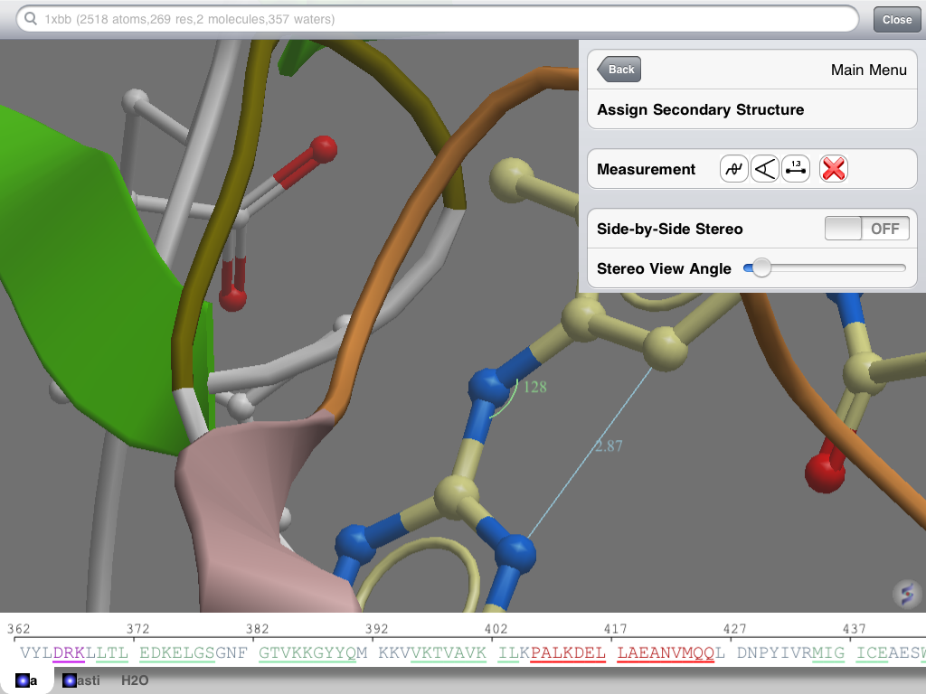 Distances and Angles
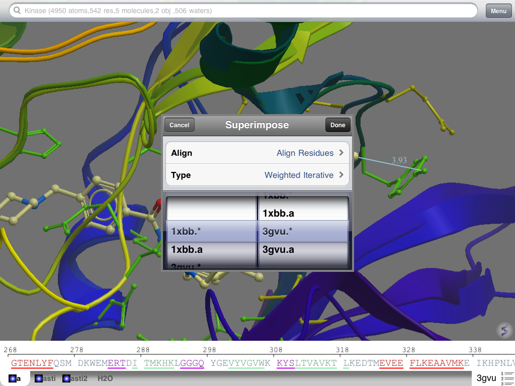 New in v1.5 Superimpose Protein Structures
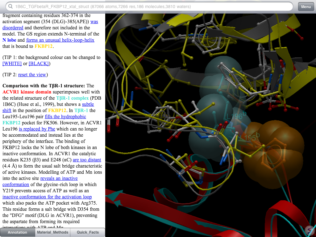 Fully Interactive Molecular Documents
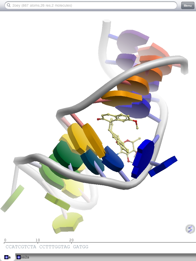 Browse protein, DNA, and drug molecules in 3D on the iPhone or iPad
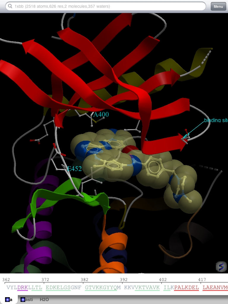 Customizable Representations
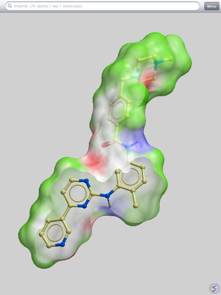 Display molecule surfaces
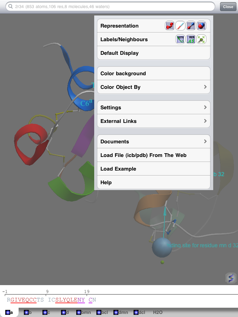 iMolview Menu Options
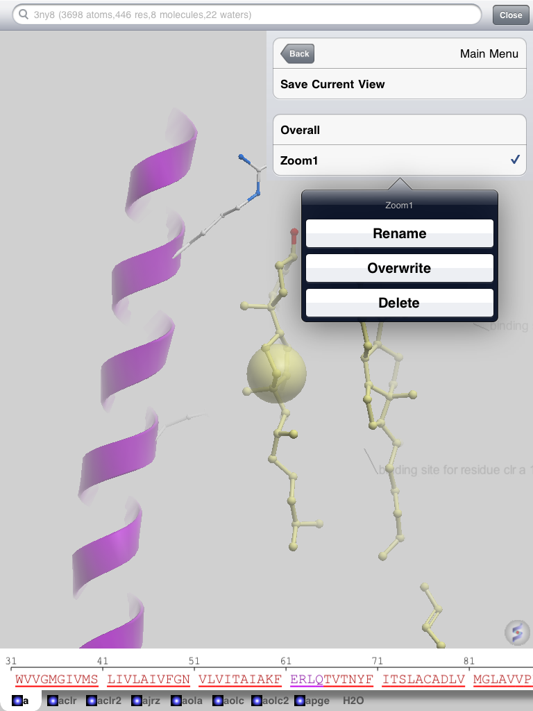 Slide Menu (v1.4)
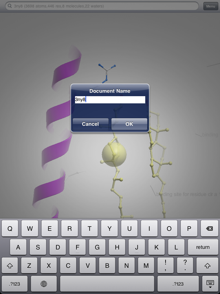 Save Documents (v1.4)
 Click to enlarge image.