[ Structures Used | Load Structures | Superimpose Receptors | Extract and Align Sequences | Select Ligand Neighbors | Compare Sequence Conservation | Receptor Surface | Ligand Contacts | UniProt Alignment ]
Compare receptor binding pockets using structural superposition, sequence alignment, ligand-neighbor selection, and molecular surfaces. The example uses a GLP-1R small-molecule complex together with retatrutide-bound GCGR and GIPR structures.
| Video tutorial: ICM Analysis of Triple Agonist Binding Pockets. |
The following PDB entries are used:
- 6XOX: GLP-1R with the small-molecule agonist LY3502970 (orforglipron; PDB ligand V6G). This structure provides the detailed ligand-pocket example.
- 8YW5: GCGR with the peptide agonist retatrutide.
- 8YW4: GIPR with the peptide agonist retatrutide.
- 8YW3: GLP-1R with retatrutide. This structure appears on the introductory slide but is not loaded in the illustrated comparison.
Important: Because 6XOX contains a small-molecule agonist whereas 8YW5 and 8YW4 contain a peptide, the analysis illustrates differences in aligned receptor positions and pocket geometry. For a matched-ligand comparison of retatrutide across all three receptors, include 8YW3.
| Structures used to compare GLP-1R, GCGR and GIPR. Video 00:00. |
Load the three structures from the Protein Data Bank.
- Select PDB Search from the ICM Search toolbar and enter 6XOX. Load the structure.
- Expand the 6XOX object in the Workspace Panel. Identify receptor chain r (GLP1R_HUMAN) and ligand entry rv6g. Other chains include associated signaling or stabilizing proteins.
- Repeat the PDB search for 8YW5 and 8YW4. In the example objects, the receptor is chain r, and the retatrutide peptide is chain p.
- Use the visibility controls in the Workspace Panel to display only the chains required for the comparison. Hidden chains remain available for later inspection.
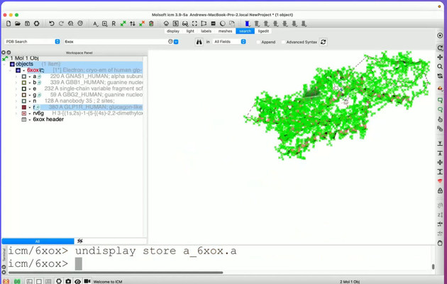
| Loading the 6XOX structure using PDB Search. Video 02:07. |
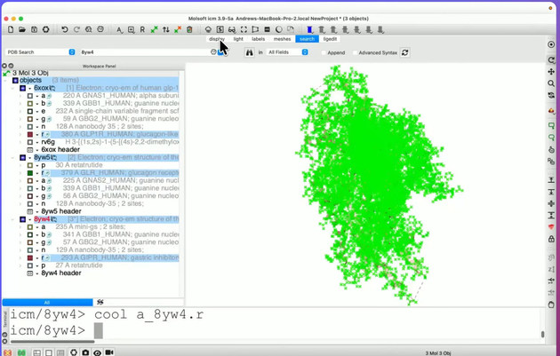
| The three receptor complexes in the ICM Workspace Panel. Video 03:22. |
Chain names are those shown in the example structures; they may differ for other PDB entries or assemblies.
Superimpose the receptors to compare their transmembrane helices and extracellular binding regions in a common coordinate frame.
- Select receptor chains 6xox.r, 8yw5.r, and 8yw4.r in the Workspace Panel. Exclude G proteins and ligand peptides from the fitting selection.
- Use the ICM structure superposition tools to fit the receptor chains, concentrating on the conserved transmembrane core.
- Display the receptors using different colors and inspect the alignment of the transmembrane helices.
- Show the bound ligands to identify the extracellular binding regions, and rotate or zoom the view to compare their positions.
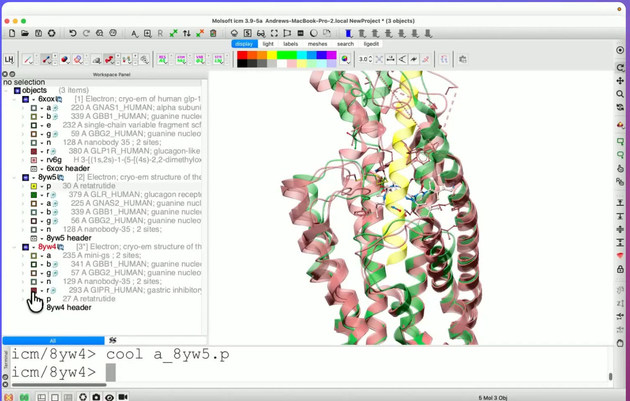
| Superimposed GPCR structures. Video 03:40. |
For structures with missing loops or different extracellular-domain coverage, assess the quality of the transmembrane-core superposition before comparing individual side chains.
The structure-linked alignment allows corresponding amino acids in GLP-1R, GCGR, and GIPR to be identified and selected in the molecular view.
- Select the three receptor chains in the Workspace Panel.
- Use Extract Sequences to Table... from the receptor context menu to create sequence records for the structures.
- Align the extracted sequences to create a three-way sequence alignment. In this example the alignment is named aln.
- Display the alignment below the 3D window and enable Sync with workspace where available, so selected residues can be located in the corresponding structural objects.
- Scroll through the alignment to identify conserved positions, substitutions, and gaps in the modeled receptor regions.
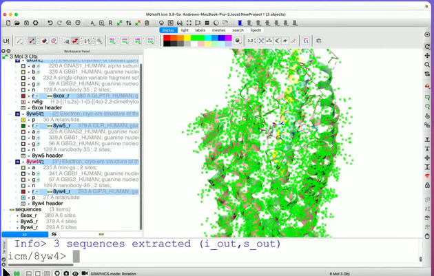
| Extracting receptor sequences. Video 04:01. |
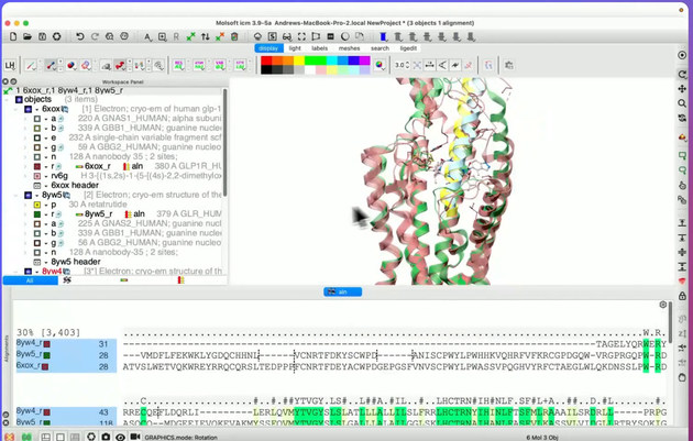
| Three-way alignment linked to the 3D structures. Video 04:12. |
The example structures contain different numbers of modeled residues; gaps in structure-derived sequences do not necessarily represent deletions in the full-length proteins.
A distance-based selection identifies residues near the ligand in the reference receptor. The example uses a 5 Å cutoff.
- Display the bound ligand in the receptor selected as the reference pocket, such as the small molecule in 6XOX.
- Select the ligand atoms and open Select Neighbors.
- Set Radius = 5 Å. The dialog provides options such as same_object, other_objects, and all_objects to control the scope of the selection.
- Apply the selection and inspect the highlighted receptor atoms and side chains in the 3D window.
- Use the linked alignment to inspect the corresponding residue positions in GCGR and GIPR.
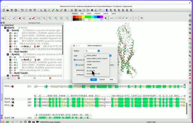
| Select Neighbors dialog with a 5 Å radius. Video 04:52. |
Note: The 5 Å cutoff identifies nearby atoms geometrically; it does not establish a direct energetic interaction. When multiple superimposed receptors are displayed, the selection scope determines which objects contribute to the selected neighborhood.
Use the alignment to identify which of the selected binding-site positions are conserved and which differ between GLP-1R, GCGR, and GIPR.
- In the alignment display settings, choose consensus-strength coloring to highlight conserved columns.
- Use Propagate to ALL seq. to extend a selection to corresponding positions in all three aligned sequences, and Highlight Selection to show the selection clearly.
- Inspect each selected position in the sequence alignment and the 3D receptor models. Compare amino-acid identity, chemical properties, and side-chain orientation.
- Use the alignment to determine corresponding residue numbers rather than matching residue numbers across the different PDB structures directly.
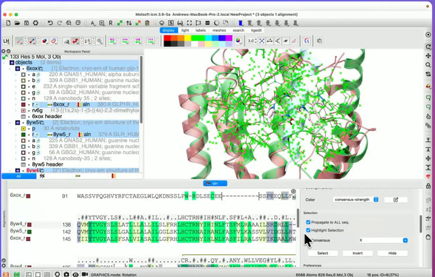
| Alignment controls for selection and conservation coloring. Video 05:37. |
Residues aligned in sequence may still differ in orientation or accessibility because of local receptor conformations, helix movements, or ligand-dependent effects.
A molecular surface can show where the selected residues are exposed around the ligand-binding pocket.
- Return to the GLP-1R structure and display its molecular skin surface using Molecular Surfaces in the receptor context menu. The example surface is named g_skin_6xox_r.
- Hide unnecessary representations and orient the model so the ligand-binding cavity is visible.
- Use the Meshes controls to adjust the surface display.
- Choose By Atom Selection to paint the relevant surface area. In the example, green is selected with BrushSize = 4.
- Rotate the receptor and inspect which selected residues contribute to the pocket surface.
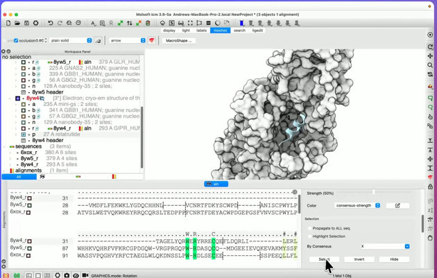
| Molecular-skin surface of GLP-1R. Video 09:06. |
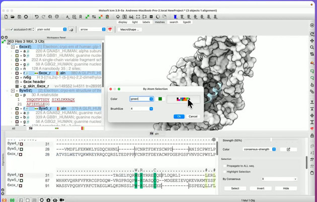
| Color selected surface regions by atom selection. Video 09:33. |
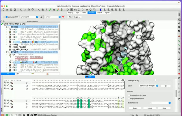
| Highlighted surface regions around the receptor pocket. Video 09:53. |
Note: The green surface patches indicate selected regions painted in the viewer. They do not represent a calculated binding-energy or sequence-conservation scale.
Display the bound small molecule and nearby receptor side chains to examine the detailed pocket environment in 6XOX.
- Temporarily hide the molecular surface. Display the GLP-1R cartoon, the ligand as sticks, and the neighboring receptor side chains.
- Enable residue labels. Examples visible in the recording include Y220, W33, Y205, C226, T298, M233, and K197.
- Select each residue of interest and inspect the corresponding aligned positions in the GCGR and GIPR structures.
- Compare side-chain identity, size, polarity, and orientation to identify potential receptor-specific binding-pocket features.
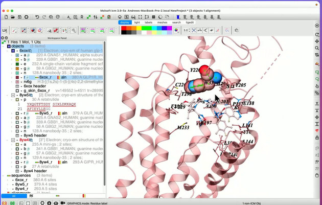
| GLP-1R side-chain labels displayed around the bound agonist. Video 11:16. |
Residue labels refer to the 6XOX structure. Use the sequence alignment to translate positions between receptors; corresponding residue numbers should not be assumed to be identical. Structural differences alone do not establish differences in potency or selectivity.
Full-length reference sequences help identify residues absent from the experimental structures and establish a consistent residue-numbering reference.
- Select File/Load/UniProt... and enter the appropriate receptor UniProt ID.
- Load the full-length receptor sequences. In the example they are labeled GLP1R_HUMAN, GIPR_HUMAN, and GLR_HUMAN (glucagon receptor).
- Create a separate alignment that includes the full-length receptor sequences and the structure-derived GLP-1R sequence. The example uses alignment tab aln_1.
- Inspect the N-terminal region and other gaps to distinguish residues absent from a structure from differences in the full-length proteins.
- Use the sequence alignment context menu and choose Draw Box to mark residue blocks of interest.
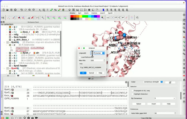
| Loading receptor sequences from UniProt. Video 12:54. |
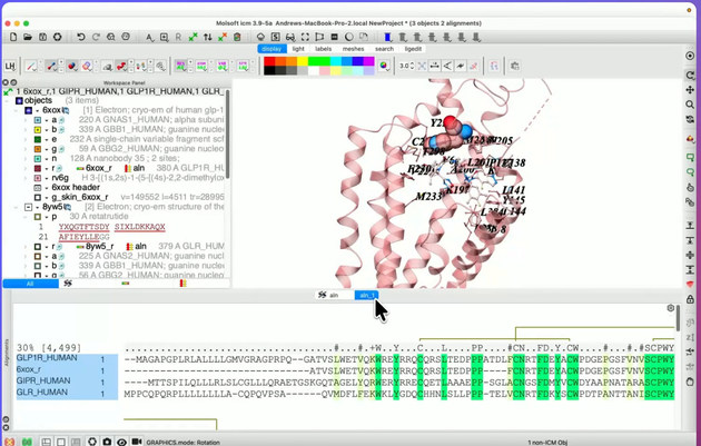
| Full-length reference sequence alignment. Video 14:12. |
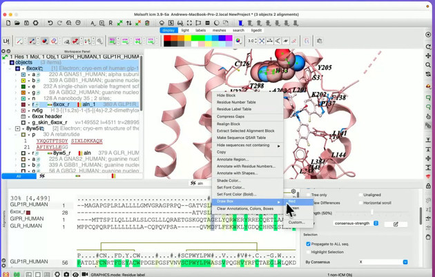
| Draw Box option for annotation in the alignment. Video 15:30. |